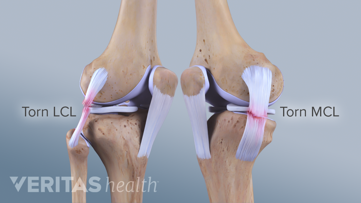
2 Ligaments In The Knee - Knee Ligaments Pic Back In Action - This means there have been small tears in the ligament.. Injury occurs when stress or force is applied to the outside of the knee, when it is still bent or pivoting. Tendons connect the knee bones to the leg muscles that move the knee joint. As a hinge joint, the knee is meant only to move in one direction. There is major pain, tenderness and swelling on the inner side of the knee. Anterior cruciate ligament (acl) is the most commonly injured knee ligament.
Anterior cruciate ligament (acl) is the most commonly injured knee ligament. The knee joint is a hinge type synovial joint, which mainly allows for flexion and extension (and a small degree of medial and lateral rotation). The medial collateral ligament (mcl) is one of the four ligaments that are critical to maintaining the mechanical stability of the knee joint. Its function is to prevent damage to the joint if adverse forces hit the inner part. These ligaments help stabilize the knee.
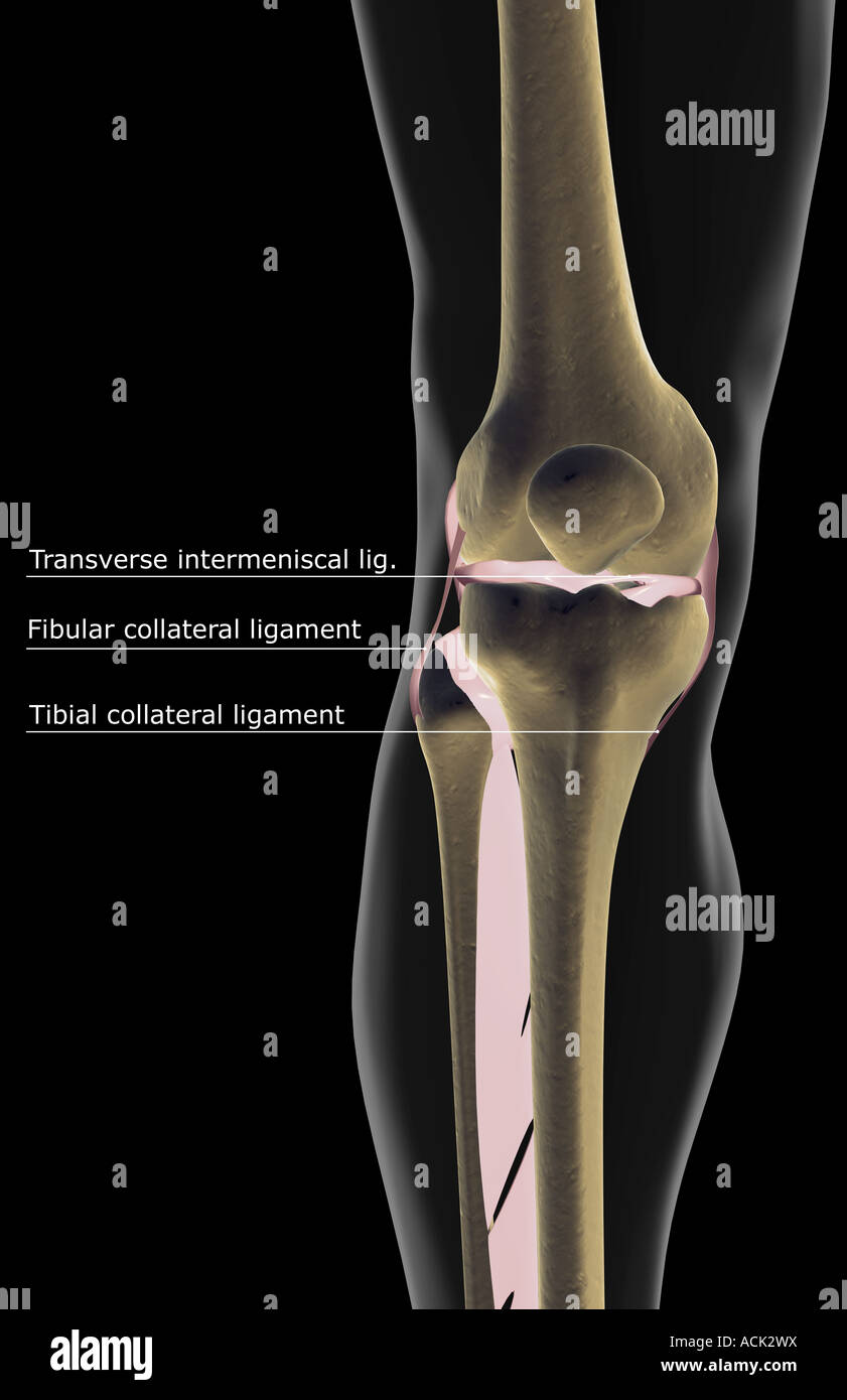
Human Knee Ligaments High Resolution Stock Photography And Images Alamy from c8.alamy.com The acl is the most commonly known knee ligament injury, and also the most common in occurrence. Posterior cruciate ligament (pcl) also links the thigh bone to the shin. There are four main ligaments that stabilize the knee that contain or restrain motion between the thigh bone and leg bone: The medial meniscus is attached medially to the tibial/medial collateral ligament and to the capsule of the joint of the knee, while the lateral meniscus is attached to neither. It's located deep inside the knee and in front of the posterior cruciate ligament. These ligaments connect the femur and tibia, holding them in place, providing stability, and preventing dislocation. It has been slightly stretched, but is still able to help keep the knee joint stable. Situated on the outer part of the knee, it is located between the fibula and femur.
Grade 2 — noticeable looseness in the knee (the knee opens up about 5 millimeters) when moved by hand.
As a hinge joint, the knee is meant only to move in one direction. There is major pain, tenderness and swelling on the inner side of the knee. Two of these ligaments are in the center of the joint, and they cross each other. A knee ligament injury is a sprain of one or more of the four ligaments in the knee, either the medial collateral ligament (mcl), lateral collateral ligament (lcl), posterior cruciate ligament (pcl), or the anterior cruciate ligament (acl). This means there have been larger tears in the ligament, but it is not completely torn. There are two cruciate ligaments, anterior (acl) and posterior (pcl). The ligament is mildly damaged in a grade 1 sprain. It connects the thigh bone to the shin bone. The medial meniscus is attached medially to the tibial/medial collateral ligament and to the capsule of the joint of the knee, while the lateral meniscus is attached to neither. The knee has four main ligaments: They cross over each other, hence their name, and resemble the st andrews cross (x). The main ligament is called the lateral collateral ligament. The knee joint, for instance, has four major ligaments, one on each side of the knee and two that run diagonally across the front and back of the kneecap.
The ligament is mildly damaged in a grade 1 sprain. There are two cruciate ligaments, anterior (acl) and posterior (pcl). The medial meniscus is attached medially to the tibial/medial collateral ligament and to the capsule of the joint of the knee, while the lateral meniscus is attached to neither. The result in grade ii sprains of the lateral ligament compartment (lat) was generally good, although residual lateral laxity was common. This causes the lateral meniscus to become more mobile (and sadly more easy to tear).
Lateral Collateral Ligament Lcl Injuries from embed.widencdn.net The four main ligaments in the knee connect the femur (thighbone) to the tibia (shin bone), and include the following: The cruciates are the most important knee ligaments in providing stability of the knee. The knee joint, for instance, has four major ligaments, one on each side of the knee and two that run diagonally across the front and back of the kneecap. It's located deep inside the knee and in front of the posterior cruciate ligament. This causes the lateral meniscus to become more mobile (and sadly more easy to tear). The ligaments of the knee joint can be divided into two groups; The knee joint is a hinge type synovial joint, which mainly allows for flexion and extension (and a small degree of medial and lateral rotation). The ligament, located in the center of the knee, that controls rotation and forward movement of the tibia (shin bone).
The anterior cruciate ligament, the posterior cruciate ligament, the medial collateral ligament, and the lateral collateral ligament.
Tendons connect the knee bones to the leg muscles that move the knee joint. These ligaments connect the femur and tibia, holding them in place, providing stability, and preventing dislocation. The medial collateral ligament (mcl) is one of the four ligaments that are critical to maintaining the mechanical stability of the knee joint. Injury occurs when stress or force is applied to the outside of the knee, when it is still bent or pivoting. These ligaments help stabilize the knee. A knee ligament injury is a sprain of one or more of the four ligaments in the knee, either the medial collateral ligament (mcl), lateral collateral ligament (lcl), posterior cruciate ligament (pcl), or the anterior cruciate ligament (acl). The ligamentous sleeve spans the entire medial side of the knee from the medial aspect of the extensor mechanism to the posterior aspect of the knee Injuries to the medial collateral ligament most often happen when the knee is hit directly on its outer side. There are two cruciate ligaments, anterior (acl) and posterior (pcl). In grade iii sprains, the results were much worse, with a high frequency of persisting severe or gross lateral laxity, insufficiency of the acl, muscle weakness, and posttraumatic osteoarthritis of the. The anterior cruciate ligament, the posterior cruciate ligament, the medial collateral ligament, and the lateral collateral ligament. There is major pain, tenderness and swelling on the inner side of the knee. The ligament, located in the center of the knee, that controls rotation and forward movement of the tibia (shin bone).
A knee ligament injury is a sprain of one or more of the four ligaments in the knee, either the medial collateral ligament (mcl), lateral collateral ligament (lcl), posterior cruciate ligament (pcl), or the anterior cruciate ligament (acl). These ligaments, which cross each other to form an x shape, connect the femur to the tibia. It connects the thigh bone to the shin bone. Tendons connect the knee bones to the leg muscles that move the knee joint. There are four main ligaments that stabilize the knee that contain or restrain motion between the thigh bone and leg bone:
Arcuate Ligament Proscan Education from info.mrionline.com The anterior cruciate ligament, the posterior cruciate ligament, the medial collateral ligament, and the lateral collateral ligament. The ligaments of the knee joint can be divided into two groups; Tendons connect the knee bones to the leg muscles that move the knee joint. There are four main ligaments that stabilize the knee that contain or restrain motion between the thigh bone and leg bone: This knee ligament runs diagonally through the center of the knee and supports and stabilizes the meeting place of the tibia, femur, and patella (the kneecap). This means there have been larger tears in the ligament, but it is not completely torn. The medial meniscus is attached medially to the tibial/medial collateral ligament and to the capsule of the joint of the knee, while the lateral meniscus is attached to neither. The four main ligaments in the knee connect the femur (thighbone) to the tibia (shin bone), and include the following:
The result in grade ii sprains of the lateral ligament compartment (lat) was generally good, although residual lateral laxity was common.
The main ligament is called the lateral collateral ligament. Posterior cruciate ligament (pcl) also links the thigh bone to the shin. Knee sprains are named for the specific ligament that has been torn or injured: The anterior cruciate ligament, or acl, is located slightly in front of the posterior cruciate ligament, or pcl. The cruciate ligaments are two strong, short ligaments located in the center of the knee joint. They sit deep inside the middle of the joint attaching to the tibia and femur. The knee has four main ligaments: The anterior cruciate ligament, the posterior cruciate ligament, the medial collateral ligament, and the lateral collateral ligament. The four ligaments joint work together to support and stabilize it as it moves about. Situated on the outer part of the knee, it is located between the fibula and femur. It's located deep inside the knee and in front of the posterior cruciate ligament. Injured ligaments are considered sprains and are graded on a severity scale. The knee joint, for instance, has four major ligaments, one on each side of the knee and two that run diagonally across the front and back of the kneecap.
Injured ligaments are considered sprains and are graded on a severity scale 2 liga. Anterior cruciate ligament (acl) is the most commonly injured knee ligament.

/image15.jpg?width=900&height=806&name=image15.jpg)


0 Komentar